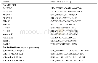
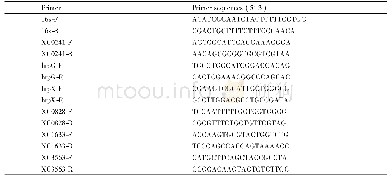
《Table 1 Primary antibodies used in this study》
 提示:宽带有限、当前游客访问压缩模式
本系列图表出处文件名:随高清版一同展现
《Effect of sevoflurane preconditioning on astrocytic dynamics and neural network formation after cerebral ischemia and reperfusion in rats》
Iba-1:Ionized calcium binding adaptor molecule-1;GFAP:glial fibrillary acidic protein;MAP2:microtubule associated protein 2;DCX:doublecortin;NF-70:neurofilament-70 kDa;DAPI:4',6-diamidino-2-phenylindole.
Brain sections were washed in 0.1 M PBS for 15 minutes and incubated with blocking solution(0.2%Triton X-100and 10%donkey serum in 0.1 M PBS)for 60 minutes at room temperature.Sections were incubated with primary antibodies(Table 1)in dilute solution(0.2%Triton X-100and 5%goat serum in 0.1 M PBS)overnight at 4°C,and then incubated with secondary antibodies(1:1000;Jackson,West Grove,PA,USA)for 2 hours at room temperature.Glial fibrillary acidic protein(GFAP)was used to label astrocytes,ionized calcium binding adaptor molecule-1(Iba-1)for microglial cells,microtubule-associated protein 2(MAP2)for neurons,doublecortin(DCX)for neuroblasts,NF-70 for neurofilaments and Ki67 for cell proliferation,respectively.Nuclei were counterstained by 4′,6-diamidino-2-phenylindole(DAPI).All images were scanned with Leica SP8(Leica Microsystems Inc.,Buffalo Grove,IL,USA)or Zeiss LSM710(Carl Zeiss MicroImaging GmbH,Jena,Germany)confocal microscopes with Z-stacks.The stitching pictures were constructed by 20μm-thick Z-stacks(5μm per step)with20×oil immersion.All the image stacks were processed by ImageJ software and Adobe Photoshop CS6(Adobe Systems Inc.,San Jose,CA,USA).Nuclear morphology was used to identify the boundary of the infarct with dotted lines(Clarke,1990).The cellular nuclei of non-infarct present as normal or pyknotic(early-stage apoptosis).The nuclei of the infarct present with karyorrhexis,which indicate that the dying cells are undergoing necrosis and late-stage apoptosis.Given that all the neural nuclei can be dyed with DAPI,the microglial cells with DAPI+debris engulfment were used to assist the delineation of the infarcted edge(Li et al.,2017).At each time point,there were eight rats in each group.
| 图表编号 | XD0040608300 严禁用于非法目的 |
|---|---|
| 绘制时间 | 2019.02.01 |
| 作者 | Qiong Yu、Li Li、Wei-Min Liang |
| 绘制单位 | Department of Anesthesiology, Huashan Hospital, Fudan University、Department of Anesthesiology, Huashan Hospital, Fudan University、Department of Anesthesiology, Huashan Hospital, Fudan University |
| 更多格式 | 高清、无水印(增值服务) |


