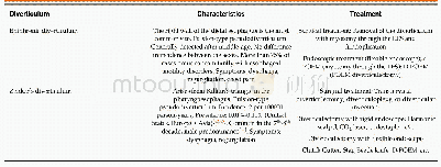
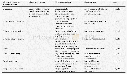
《Table 1 Summary of the characteristics of epiphrenic and Zenker’s diverticula and their treatment o
 提示:宽带有限、当前游客访问压缩模式
本系列图表出处文件名:随高清版一同展现
《Esophageal diverticulum: New perspectives in the era of minimally invasive endoscopic treatment》
LES:Lower esophageal sphincter;POEM:Peroral endoscopic myotomy;s-POEM:Salvage peroral endoscopic myotomy;D-POEM:Diverticular peroral endoscopic myotomy.
Recent studies have reported that more than 75%of epiphrenic diverticula occur concomitantly with esophageal motility disorders[2-8].Therefore,evaluation of esophageal motility using high resolution manometry(HRM)is recommended before deciding on the intervention(Figure 1B)[9,10].The right esophageal wall is more susceptible to epiphrenic diverticula[2,11],although the reason for this is still unclear.In contrast,spontaneous esophageal rupture tends to occur through the left wall(Table1)[12].
| 图表编号 | XD0064274600 严禁用于非法目的 |
|---|---|
| 绘制时间 | 2019.03.28 |
| 作者 | Hiroki Sato、Manabu Takeuchi、Satoru Hashimoto、Ken-ichi Mizuno、Koichi Furukawa、Akito Sato、Junji Yokoyama、Shuji Terai |
| 绘制单位 | Division of Gastroenterology, Niigata University Medical and Dental Hospital、Division of Gastroenterology, Nagaoka Red Cross Hospital、Division of Gastroenterology, Niigata University Medical and Dental Hospital、Division of Gastroenterology, Niigata Univer |
| 更多格式 | 高清、无水印(增值服务) |
