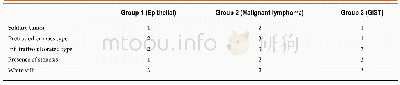
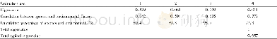
《Table 4 Summary of the endoscopic characteristics of the small intestinal tumors》
 提示:宽带有限、当前游客访问压缩模式
本系列图表出处文件名:随高清版一同展现
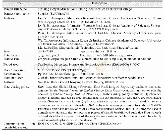
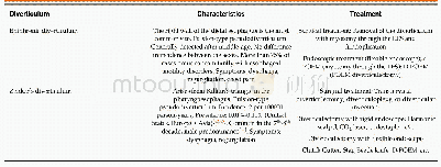
《Endoscopic characteristics of small intestinal malignant tumors observed by balloon-assisted enteroscopy》
1:More likely;2:Intermediate;3:Less likely;GIST:Gastrointestinal stromal tumor.
In the current study,we described the endoscopic characteristics of tumor location and morphology of small intestinal malignant tumors.Small intestinal malignant tumors were classified into three groups,and statistical analyses were performed between the groups.Patients’clinical background parameters including age,symptoms,and laboratory data were not significantly different between the groups.First,we evaluated the endoscopic characteristics of tumor location and the number of tumors.Approximately three quarters of epithelial tumors were found in the duodenum or jejunum,and all were observed as solitary lesions(Table 2).Previous studies using BAE reported that primary small intestinal adenocarcinoma was located mainly in the duodenum or jejunum with a range of 77.8%–100.0%[6,7,11-14].Primary small intestinal adenocarcinoma was reported as a solitary lesion in several studies[6,7,12-14],which was consistent with our results,whereas metastatic tumors were sometimes observed as multiple lesions[11,15].In our classification,primary and metastatic tumors were classified into the same groups;however,it might be better to distinguish between metastatic and primary tumors.In the current study,malignant lymphoma lesions were located mainly in the jejunum and ileum,and approximately60%were multiple lesions(Table 2),consistent with previous reports[11-14,16].GISTs are reported mainly as solitary jejunal tumors[11-13,17,18].Nakano et al[19]reported that 76%of patients with GIST had jejunal lesions,and that 3/25 patients had tumors in multiple sites(stomach and jejunum:1;duodenum and jejunum:1;and stomach,duodenum,and jejunum:1).A particular type of GIST that is associated with neurofibromatosis type1 appears as multiple tumors[20-22].However,as our results,most GISTs appeared as a solitary jejunal tumor,except for neurofibromatosis type1 associated type[20-22].
| 图表编号 | XD0058775600 严禁用于非法目的 |
|---|---|
| 绘制时间 | 2019.05.16 |
| 作者 | Tomofumi Horie、Naoki Hosoe、Kaoru Takabayashi、Yukie Hayashi、Kenji JL Limpias Kamiya、Ryoichi Miyanaga、Shinta Mizuno、Kayoko Fukuhara、Seiichiro Fukuhara、Makoto Naganuma、Masayuki Shimoda、Haruhiko Ogata、Takanori Kanai |
| 绘制单位 | Division of Gastroenterology and Hepatology,Department of Internal Medicine,School of Medicine,Keio University、Center for Diagnostic and Therapeutic Endoscopy,School of Medicine,Keio University、Center for Diagnostic and Therapeutic Endoscopy,School of Med |
| 更多格式 | 高清、无水印(增值服务) |