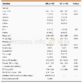
《Table 1 Demographic and clinical characteristics of the subjects》
 提示:宽带有限、当前游客访问压缩模式
本系列图表出处文件名:随高清版一同展现
《Regional gray matter abnormality in hepatic myelopathy patients after transjugular intrahepatic portosystemic shunt: a voxel-based morphometry study》
Data are expressed as the mean±SD with the exception of sex,handedness,Child-Pugh stage,and West-Haven HE grade(n).*:One-way analysis of variance;#:Chi-square test;:nonparametric Kruskal-Wallis H test;$:independent-samples t test.HE:Hepatic encephal
One-way analysis of variance across the hepatic myelopathy,non-hepatic myelopathy,and healthy control groups showed significant differences in gray matter volume in both thalami,putamina,globi pallidi,parahippocampi,and in the cerebellum and vermis(Table 2 and Figure 2).As compared with the healthy controls,both hepatic myelopathy and non-hepatic myelopathy patients showed increased gray matter volumes in both thalami and parahippocampi,with decreased gray matter volumes in the putamina,globi pallidi,cerebellum,and vermis(Table 3 and Figure 3A&B).Additionally,compared with the non-hepatic myelopathy group,gray matter volume was increased in the right cau-date nucleus,and decreased in the left insula,left thalamus,left superior frontal gyrus,and middle cingulate cortex in the hepatic myelopathy group(Table 4 and Figure 3C).
| 图表编号 | XD0040614300 严禁用于非法目的 |
|---|---|
| 绘制时间 | 2019.05.01 |
| 作者 | Kang Liu、Gang Chen、Shu-Yao Ren、Yuan-Qiang Zhu、Tian-Lei Yu、Ping Tian、Chen Li、Yi-Bin Xi、Zheng-Yu Wang、Jian-Jun Ye、Guo-Hong Han、Hong Yin |
| 绘制单位 | Department of Radiology, Xijing Hospital, Air Force Military Medical University (Fourth Military Medical University)、Department of Radiology, Lanzhou General Hospital, Lanzhou Military Command、Xijing Hospital of Digestive Diseases, Air Force Military Medi |
| 更多格式 | 高清、无水印(增值服务) |

