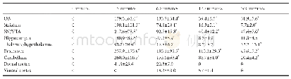
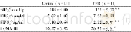
《Table 1 Recombinant human insulin concentration (pg/mg tissue) in different brain regions over a 6-
 提示:宽带有限、当前游客访问压缩模式
本系列图表出处文件名:随高清版一同展现
《Rapid transport of insulin to the brain following intranasal administration in rats》
Data represent mean±SEM from five rats in each group.*P<0.05,vs.0 minute(one-way analysis of variance followed by Bonferroni post hoc analysis).OB:Olfactory bulb;SN:subtantia nigra;VTA:ventral tegmental area.
The specificity of the ELISA kit for rh-Ins was first validated based on positive controls of human insulin and rat samples from the control group.The calculated concentration of two positive controls in each ELISA consistently reached within 5%range of predicted values.The concentration of rhIns from PBS-treated control rats were all below detectable limit,therefore they were assigned a value of 0.As shown in Table 1,significant amounts of rh-Ins were detected in all brain regions except the cerebral cortex in rh-Ins-treated rats.Rh-Ins reached the peak value at 15 minutes and declined substantially overtime,but remained significantly higher than baseline levels in most regions at 6 hours.Neither the dorsal or ventral division of the cortex showed a significant increase over baseline at any time points.
| 图表编号 | XD0040617200 严禁用于非法目的 |
|---|---|
| 绘制时间 | 2019.06.01 |
| 作者 | Lir-Wan Fan、Kathleen Carter、Abhay Bhatt、Yi Pang |
| 绘制单位 | Department of Pediatrics,University of Mississippi Medical Center、Department of Pediatrics,University of Mississippi Medical Center、Department of Pediatrics,University of Mississippi Medical Center、Department of Pediatrics,University of Mississippi Medica |
| 更多格式 | 高清、无水印(增值服务) |
![表3 重组人骨形态发生蛋白2(recombinant human bone morphogenetic protein,rhBMP-2)相关不良反应总结[34]](http://bookimg.mtoou.info/tubiao/gif/LCKY202003016_04100.gif)