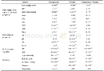
《Table 3 Anatomical locations of regions with significant clinical correlation in iRBD patients》
 提示:宽带有限、当前游客访问压缩模式
本系列图表出处文件名:随高清版一同展现
《Assessing gray matter volume in patients with idiopathic rapid eye movement sleep behavior disorder》
iRBD:Idiopathic rapid eye movement sleep behavior disorder;BA:Brodmann area;EMG:electromyographic.Multiple regression analysis,P<0.01uncorrected for multiple comparisons.
Relative to controls,iRBD patients exhibited reduced GMV in the right rolandic operculum,right postcentral gyrus,right insular lobe,right anterior cingulate gyrus,right precuneus,right posterior cingulate gyrus,left rectus gyrus,and bilateral superior frontal gyrus.Furthermore,iRBD patients displayed increased GMV in the right middle temporal gyrus and left cerebellar posterior lobe(Figure 2 and Table 2).
| 图表编号 | XD0040615300 严禁用于非法目的 |
|---|---|
| 绘制时间 | 2019.05.01 |
| 作者 | Xian-Hua Han、Xiu-Ming Li、Wei-Jun Tang、Huan Yu、Ping Wu、Jing-Jie Ge、Jian Wang、Chuan-Tao Zuo、Kuang-Yu Shi |
| 绘制单位 | PET Center, Huashan Hospital, Fudan University、PET Center, Huashan Hospital, Fudan University、Department of Radiology, Huashan Hospital, Fudan University、Department of Neurology, Huashan Hospital, Fudan University、PET Center, Huashan Hospital, Fudan Unive |
| 更多格式 | 高清、无水印(增值服务) |
查看“Table 3 Anatomical locations of regions with significant clinical correlation in iRBD patients”的人还看了
-

- Table 1 Trough values and trough separations of the opti-cal intensity changing with locations of the droplets
-

- Table 1 Difference significance of RNA absorbance of samples with and without liquid nitrogen grinding
-

- Table 1 miRNAs with significantly altered expression in retinas of mice with OIR identified by microarray
-

- Table 2 Brain regions with significant gray matter volume differences between iRBD patients and controls


