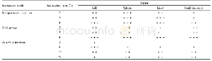
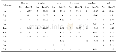
《Table 1 Detection of positive signals for pathogen infection in various organs of tilapia after art
 提示:宽带有限、当前游客访问压缩模式
本系列图表出处文件名:随高清版一同展现
《A Pathological Study of GIFT Strain of Nile Tilapia(Oreochromis niloticus) Infected by Streptococcus agalactiae》
The positive signals for pathogen infection were identified according to the presence/absence,amount and coloring degree of brownish-yellow particles under an optical microscope.Negative cells were blue.\""-\"":no positive signals;\""+\"":weak
After artificial inoculation of S.agalactiae into GIFT strain of Nile tilapia via intraperitoneal injection,oral gavage and in vitro immersion,dynamic location and distribution of positive signals for pathogen infection in the gill,spleen,liver and small intestine tissues of tilapia were analyzed.As shown in Table 1,positive signals for pathogen infection were found in four organs of tilapia at2 h post-inoculation in intraperitoneal injection and oral gavage groups.Specifically,the strongest positive signals in intraperitoneal injection group were found in the spleen,while that in oral gavage group appeared in the small intestine.In in vitro immersion group,positive signals were observed in the gill and spleen of tilapia at 5 h post-inoculation,and those in the gill were much stronger.According to the appearance time and intensity of positive signals for pathogen infection,the appearance time of positive signals in intraperitoneal injection group demonstrated an order of spleen→liver and gill→small intestine;positive signals in oral gavage group appeared in the order of small intestine→gill and spleen→liver;the appearance time of positive signals in in vitro immersion group showed an order of gill→spleen→liver and small intestine.
| 图表编号 | XD00183125300 严禁用于非法目的 |
|---|---|
| 绘制时间 | 2018.10.01 |
| 作者 | Wei LUO、Xi GAN、Jiajie ZHU、Qiuwei AO、Yun TAN、Ming CHEN、Yongju LUO |
| 绘制单位 | Guangxi Academy of Fishery Sciences、Guangxi Key Laboratory for Aquatic Genetic Breeding and Healthy Aquaculture、Guangxi Academy of Fishery Sciences、Guangxi Key Laboratory for Aquatic Genetic Breeding and Healthy Aquaculture、Guangxi Academy of Fishery Scie |
| 更多格式 | 高清、无水印(增值服务) |