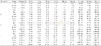
《Table 2 Average MD and retinal pigmentation in 928 observed eyes at the patients’first visit》
 提示:宽带有限、当前游客访问压缩模式
本系列图表出处文件名:随高清版一同展现
《Visual field mean deviation and relevant factors in 928 Chinese retinitis pigmentosa patients》
MD:Mean deviation.
MD indicates the mean difference between the normal expected retinal sensitivity in terms of age and visual acuity and the measured patient sensitivity[5].This definition has already indicated the comparation with normal eyes,so there is no need to set a normal comparative group when MD is used for clinical research.In this paper,as MD is the major research index,there is also no need to set normal comparative groups for minor indexes such as visual acuity,gender,age,and so on.Calculating according to the above linear regression equation of MD on visual acuity in 928 observed eyes at the patients’first visit,the authors expected that when visual acuity was lower than 1.0 the MD should be lower than-7.75 dB.As the R2 value of the equation was 0.6168,it was possible that the equation couldn’t well express the correlation between MD and visual acuity.Table 1 showed when average MD inobserved eyes was-9.18 dB,the visual acuity was still at 1.0and above.It indicated that in the early stage of RP the visual acuity of observed eyes would maintain normal.The socalled early stage in this paper was that when average MD was higher than or equal to-9.18 dB,but the authors suggested the clinical early stage of RP should be defined as the period from the onset of the disease to the time point when patient’s visual acuity was just under 1.0.This definition accorded with the pathologic process of RP.The progressive atrophy of the rod photoreceptor cells leads to the secondary death of the cones in RP,affected individuals often present with night blindness and constricted visual fields,but in the early stage of RP the central vision is normal or nearly normal.Eventually central vision is lost as the cone cells degenerate[6-9].Clinically,visual field becomes more and more narrow,accordingly visual field sensitivity or MD is lower and lower.When MD is low to a certain extent,visual acuity will be less than 1.0.
| 图表编号 | XD0023069200 严禁用于非法目的 |
|---|---|
| 绘制时间 | 2018.12.18 |
| 作者 | Hui Ye、Xiao-Ping Xia |
| 绘制单位 | Department of Ophthalmology, the Third Affiliated Hospital of Sun Yat-sen University、Department of Ophthalmology, the Third Affiliated Hospital of Sun Yat-sen University |
| 更多格式 | 高清、无水印(增值服务) |


