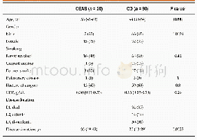
《Table 2 Cross-tabulation of the study characteristics》
 提示:宽带有限、当前游客访问压缩模式
本系列图表出处文件名:随高清版一同展现
《Role of innovative 3D printing models in the management of hepatobiliary malignancies》
Both student education in liver anatomy and resident training in liver surgical techniques with the use of 3D printed models are the second most widely studied application.Ten studies refer to this field,five of which were designed as case reports or case series[28,32,37,38,41],one was an observational cohort study[34]and four were RCTs[35,36,39,40].The case reports and case series state that 3D printed models could help in both the understanding of liver anatomy and surgical training,but fail to provide objective evidence.Dhir et al[33]was the first to use 3D printed liver models to train a cohort of 15 individuals in EUS-guided biliary drainage(EUS-BD)and,moreover,to measure the educational outcomes.The success rates were:100%for needle puncture,82.35%for wire manipulation and 80%for stent placement.However,the more experienced trainees were found to score lower in stent placement.This may be attributed to the small sample size that may not have supported safe comparison tests.Two RCTs were published in 2016[35,36],and two in 2018[39,40],comparing the educational impact of 3D printing to more conventional strategies.Kong et al[34]randomized 61 medical students into three cohorts:the 1st was trained in 3D printed models,the 2nd in 3D virtual models on computers and the 3rd in traditional anatomy atlases.The results favored both 3D printed and virtual models against the traditional atlases.However,the sample was small,and there was no cost analysis to the use of3D printing against 3D virtual methods.In another study that same year,Kong et al[35]randomized 92 students into four groups,and trained them in different settings:The1st was trained on a 3D printed model with hepatic segments without parenchyma,the2nd on a 3D printed model with hepatic segments with transparent parenchyma,the 3rd on a biliary tract model with segmental partitions and the 4th on traditional anatomic atlases.In general,the 3rd group was found to have better results,although the samples in the randomized groups may also lack enough statistical power.Li et al[38]tried to use 3D printed models against virtual,computer-based 3D models in order to train 20 individuals in choledochoscopy.Although the randomized sample was also small,the design was sophisticated,and the results showed 3D printed models to be superior.Yang et al[39]evaluated the educational impact of 3D printed models against3D virtual models and CT-based tools,and found them to be superior.These studies provide significant evidence that 3D printing could be applied in education and training in liver surgery,although they may have some methodological weaknesses.
| 图表编号 | XD0077757900 严禁用于非法目的 |
|---|---|
| 绘制时间 | 2019.07.27 |
| 作者 | Peter Bangeas、Vassilios Tsioukas、Vasileios N Papadopoulos、Georgios Tsoulfas |
| 绘制单位 | Department of Surgery,Aristotle University of Thessaloniki、Department of School of Rural and Surveying Engineering,Aristotle University of Thessaloniki、Department of Surgery,Aristotle University of Thessaloniki、Department of Surgery,Aristotle University o |
| 更多格式 | 高清、无水印(增值服务) |


