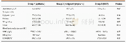
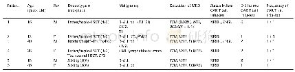
《Table 1 Patients’characteristics》
 提示:宽带有限、当前游客访问压缩模式
本系列图表出处文件名:随高清版一同展现
《Endoscopic characteristics of small intestinal malignant tumors observed by balloon-assisted enteroscopy》
N/A:Not applicable:GIST:Gastrointestinal stromal tumor;WBC:White blood cell count;Hb:Hemoglobin;LDH:Lactate dehydrogenase.
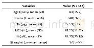
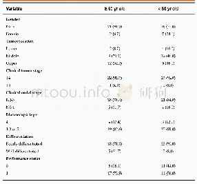
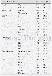
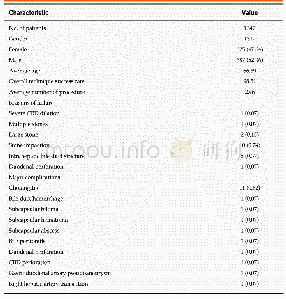
A flow diagram of patient enrollment is shown in Figure 1.In total,1328 BAE procedures were performed from March 2005 to February 2017.Of these 1328procedures,the number of patients in each group was 16(Group 1),22(Group 2),and6(Group 3)(Figure 1) .Table 1 shows the patients’characteristics.We found no statistically-significant difference in age,symptoms(epigastric pain,melena,weight loss),and blood test results(white blood cell count,hemoglobin,lactate dehydrogenase)between the groups.The endoscopic characteristics of the small intestinal malignant tumors are shown in Table 2 and Figure 2.Seventy-five percent of epithelial tumors(Group 1)were located in the upper small intestine(duodenum and jejunum),and approximately 70%of GISTs were located in the jejunum.The percentage of solitary tumors was 100%,45.5%,and 100%in Group 1,2,and 3,respectively(P<0.001).Solitary protruding or mass-type tumors were not seen in malignant lymphoma(Group 2)(P<0.001) .Solitary infiltrative ulcerated type tumors were seen only in Group1(P=0.007)(Figure 2A) .Multiple lesions with ulcerated surfaces or polyposis were seen only in Group 2,and stenosis was seen more frequently in Group 1,(68.8%,27.3%,and 0%;Group 1,2,and 3,respectively;P=0.004) .Although the difference was not statistically significant,Group1 tended to have more bleeding compared with Group 2 and 3.Enlarged white villi inside and/or surrounding the tumor were seen in 12.5%,54.5%,and 0%in Group 1,2 and 3,respectively(P<0.001)(Figure 2B and C) .We further investigated the pathological and morphological features of white villi in Group 2.Adequate biopsy samples were not obtained from four patients;therefore,we excluded data for these patients from the analysis(Table 3).Of the 22 Group 2 patients,enlarged white villi were seen in 12patients.At the biopsy sites where most of the white villi were seen,lymphoma cells infiltrated into the villi with an intact epithelium;villi were filled with lymphoma cells(Table 3 and Figure 3).When the intact epithelium was ulcerated or lymphoma cells were present in the deep mucosa,white villi could not be seen(Table 3 and Figure 4).
| 图表编号 | XD0058774900 严禁用于非法目的 |
|---|---|
| 绘制时间 | 2019.05.16 |
| 作者 | Tomofumi Horie、Naoki Hosoe、Kaoru Takabayashi、Yukie Hayashi、Kenji JL Limpias Kamiya、Ryoichi Miyanaga、Shinta Mizuno、Kayoko Fukuhara、Seiichiro Fukuhara、Makoto Naganuma、Masayuki Shimoda、Haruhiko Ogata、Takanori Kanai |
| 绘制单位 | Division of Gastroenterology and Hepatology,Department of Internal Medicine,School of Medicine,Keio University、Center for Diagnostic and Therapeutic Endoscopy,School of Medicine,Keio University、Center for Diagnostic and Therapeutic Endoscopy,School of Med |
| 更多格式 | 高清、无水印(增值服务) |