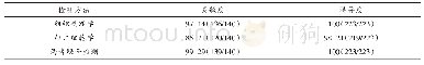
《表3 各组大鼠肠道细菌移位阳性率比较[n=5, 只 (%) ]Tab 3 Comparison of positive rates of bacterial trans-location in ra
![《表3 各组大鼠肠道细菌移位阳性率比较[n=5, 只 (%) ]Tab 3 Comparison of positive rates of bacterial trans-location in ra](http://bookimg.mtoou.info/tubiao/gif/ZGYA201902013_11000.gif) 提示:宽带有限、当前游客访问压缩模式
提示:宽带有限、当前游客访问压缩模式
本系列图表出处文件名:随高清版一同展现
《内毒素亲和吸附剂SPV对失血性休克模型大鼠肠壁通透性与细菌移位的影响》
注:与同时间点正常组比较,*P<0.05;与同时间点休克组比较,#P<0.05;与同时间点SPV低剂量组比较,ΔP<0.05Note:vs.normal group at the same time point,*P<0.05;vs.shock group at the same time point,#P<0.05;vs.SPV low-dose group at the same time point,ΔP<0.05
与同时间点正常组比较,休克组大鼠各时间点肠道细菌移位阳性率均显著上升,差异均有统计学意义(P<0.05)。与同时间点休克组比较,SPV低剂量组大鼠4~16 h各时间点以及SPV中、高剂量组大鼠1~16 h各时间点的细菌移位阳性率均显著降低,且SPV中、高剂量组大鼠1~16 h各时间点的细菌移位阳性率均显著低于SPV低剂量组,差异均有统计学意义(P<0.05);而SPV中、高剂量组大鼠同时间点肠道细菌移位阳性率比较,差异均无统计学意义(P>0.05),详见表3。
| 图表编号 | XD0047813800 严禁用于非法目的 |
|---|---|
| 绘制时间 | 2019.01.30 |
| 作者 | 刘海 |
| 绘制单位 | 唐山市开滦总医院麻醉科 |
| 更多格式 | 高清、无水印(增值服务) |
![表2 各组大鼠器官细菌移位情况比较[n(%)]](http://bookimg.mtoou.info/tubiao/gif/ZYJZ201911001_02300.gif)
