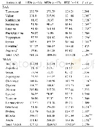
《Table 5 Comparison of progression-free survival among groups created based on regression change and
 提示:宽带有限、当前游客访问压缩模式
本系列图表出处文件名:随高清版一同展现
《Unnecessity of lymph node regression evaluation for predicting gastric adenocarcinoma outcome after neoadjuvant chemotherapy》
1Groups A+B vs Groups C+D.A:True negative lymph nodes(LNs)with no evidence of a preoperative therapy effect;B:No residual metastasis but presence of regression change in LNs;C:Residual metastasis with regression change in LNs;D:Metastasis with minimal or
All of the examined LN specimens were embedded in paraffin,and four micrometer tissue sections were stained with hematoxylin and eosin.The median number of resected LNs was 24(3-58)per case.The histopathological evidence of regression change in LNs was defined as the presence of fibrosis,aggregation of foamy histocytes or accumulation of mucin pools in LN parenchyma[6],which is shown in Figure 1.We classified tumor regression and residual tumor in LNs into four groups:A,true negative LNs with no evidence of a preoperative therapy effect;B,no residual metastasis but presence of regression change in LNs;C,residual metastasis with regression change in LNs;and D,metastasis with minimal or no regression change in LNs(Table 1 and Figure 2).All of the sections were reviewed by three experienced pathologists(Zhu YL,Yue JY and Xue LY).For a controversial diagnosis,three pathologists reviewed the sections on a multi-headed microscope until reaching an agreement.
| 图表编号 | XD0043289500 严禁用于非法目的 |
|---|---|
| 绘制时间 | 2019.01.15 |
| 作者 | Yue-Lu Zhu、Yong-Kun Sun、Xue-Min Xue、Jiang-Ying Yue、Lin Yang、Li-Yan Xue |
| 绘制单位 | Department of Pathology, National Cancer Center、National Clinical Research Center for Cancer、Cancer Hospital, Chinese Academy of Medical Sciences and Peking Union Medical College、Department of Medical Oncology, National Cancer Center、National Clinical Res |
| 更多格式 | 高清、无水印(增值服务) |
查看“Table 5 Comparison of progression-free survival among groups created based on regression change and residual tumor in ly”的人还看了
-

- 表3 2012~2016年芜湖市城乡幼托儿童报告传染病发病水平比较Table 3 Comparison of the incidence of infectious diseases among children in urban and


