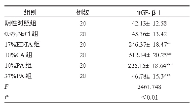
《Table 2 Protein levels (pg/mL) of TGF-β1, GDNF, bFGF and ANG1 (astrocyte secretory proteins) in the
 提示:宽带有限、当前游客访问压缩模式
本系列图表出处文件名:随高清版一同展现
《Structural and functional damage to the hippocampal neurovascular unit in diabetes-related depression》
*P<0.05,**P<0.01,vs.NVU group;#P<0.05,vs.NVU model group(NVU+G&P,NVU+glucose+corticosterone).NE:Neuron;AS:astrocyte;BM:brain microvascular endothelial cell;NVU:neurovascular unit;G&P:glucose and corticosterone;TGF-β1:tumor growth factor beta 1;GDNF:
As shown in Table 1,expression of the secretory protein LIF in brain microvascular endothelial cells was decreased in the astrocytes+brain microvascular endothelial cells group and NVU group after DD,compared with the corresponding controls(P<0.01).Moreover,LIF expression was lowest in the astrocytes+brain microvascular endothelial cells group exposed to hyperglycemia and corticosterone(P<0.01).Therefore,the secretory function of brain microvascular endothelial cells is reduced by simulated DD.Secretory function was better in the NVU co-culture system compared with the astrocytes+brain microvascular endothelial cells system.
| 图表编号 | XD0040608600 严禁用于非法目的 |
|---|---|
| 绘制时间 | 2019.02.01 |
| 作者 | Jian Liu、Yu-Hong Wang、Wei Li、Lin Liu、Hui Yang、Pan Meng、Yuan-Shan Han |
| 绘制单位 | First Hospital of Hunan University of Chinese Medicine、Hunan University of Chinese Medicine、First Hospital of Hunan University of Chinese Medicine、First Hospital of Hunan University of Chinese Medicine、First Hospital of Hunan University of Chinese Medicin |
| 更多格式 | 高清、无水印(增值服务) |


