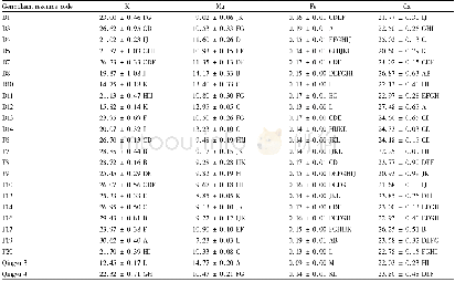
《Table 1.Bone mineral content of femur》
 提示:宽带有限、当前游客访问压缩模式
本系列图表出处文件名:随高清版一同展现
《A peptide containing the receptor binding site of insulin-like growth factor binding protein-2 enhances bone mass in ovariectomized rats》
aP<0.01 compared to sham control(ctrl)b,cP<0.05,P<0.01 compared to OVX control (ctrl)
The rats which had been ovariectomized 8 weeks prior to beginning the peptide injections consistently weighed more than the sham control rats.All groups of rats gained weight during the study;and the ovariectomized animals maintained a higher weight compared to the sham controls(Supplemental Fig.2).Analysis of areal bone mineral density(aBMD)in the femur showed that the sham animals had significantly greater aBMD compared to OVX animals at the initiation of the injection period,and this difference remained unchanged.Sham animals that received the control peptide or high-dose PEG-HBD1(data not shown)had no significant change in aBMD.All OVX groups significantly had lower aBMD(P<0.001)than the sham controls at baseline(for example 15.4%reduction)(Fig.1a) .Following 8 weeks of treatment with PEG-HBD1 peptide,the OVX high-dose peptide treatment group showed a significant increase(P<0.05)in femur aBMD compared to either their baseline value or the OVX control peptide-treated animals(Fig.1b).This change was apparent following 4 weeks of treatment and persisted for the 8 week treatment interval.Both low-and mid-dose PEG-HBD1 peptide treatment groups at 4 weeks,but only low-dose peptide treatment at 8 weeks,showed significant increases in the femoral aBMD compared to the control peptide-treated rats.When the data were expressed as percent increase over basal,the greatest response(e.g.6.2%)increase was noted in the high-dose peptidetreated animals,with the next greatest response 5.6%in the parathyroid hormone(PTH)treated group.Following intermittent exposure this PTH preparation stimulated osteoblast differentiation,which was comparable to a PTH preparation obtained for a different source(Supplemental Fig.3).The response in the OVX low-dose group(3.8%±1.5%increase,P<0.05)was significantly greater than control peptide(Fig.1c).The mid-dose group increased 3.1%±1.6%but this change was not significant.Analysis of tibial aBMD showed no significant increases at any dose.All three doses of PEG-HBD1 peptide stimulated an increase in both femoral and tibial bone mineral content(BMC)compared to the control peptide-treated groups.The percentage increase in femoral BMC varied from 3.6%to 7.2%(Table 1),and tibial BMC varied from 3.5%to 9.8%(Table 2).Sham control,sham high dose(data not shown),and OVX control peptide-treated animals showed no change.PTH increased aBMD at the femur(5.6%±2.1%,P<0.01)but not the tibia(1.4%±3.2%,p,NS)when compared to OVX control peptide responses.Analysis of the femoral BMC after 8 weeks showed significant increases in each of the treatment groups(e.g.,0.407±0.042,P<0.05,for low dose;0.404±0.038,P<0.01 for middle dose;0.418±0.044,P<0.01 for high dose;0.418±0.025,P<0.01 for PTH vs.0.390±0.010 for control peptide)(Table 1) .The high-dose treatment group response was significantly greater than the lower-dose and middle-dose groups,and it was comparable to PTH.Analysis of tibial BMC showed that the low-dose and high-dose groups had significantly greater responses,whereas the middle-dose group did not achieve significance.The response in the PTH group was comparable to the low-dose and high-dose responses(Table 2).
| 图表编号 | XD0021874200 严禁用于非法目的 |
|---|---|
| 绘制时间 | 2018.09.01 |
| 作者 | Gang Xi、Christine Wai、Clifford J.Rosen、David R.Clemmons |
| 绘制单位 | Department of Medicine, University of North Carolina at Chapel Hill、Department of Medicine, University of North Carolina at Chapel Hill、Maine Medical Center Research Institute、Department of Medicine, University of North Carolina at Chapel Hill |
| 更多格式 | 高清、无水印(增值服务) |
