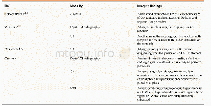
《Table 1 Imaging findings of primary gastric melanoma in the literature and our case》
 提示:宽带有限、当前游客访问压缩模式
本系列图表出处文件名:随高清版一同展现
《Primary gastric melanoma: A case report with imaging findings and 5-year follow-up》
CT:Computed tomography;MRI:Magnetic resonance imaging;GI:Gastrointestinal;T1WI:T1-weighted imaging;T2WI:T2-weighted imaging;DWI:Diffusion-weighted imaging.
Metastatic gastric melanoma are more common than primary ones;PGM are rare in clinic,and the related literature is also relatively scarce[2-5].Imaging examination is of great value in melanoma diagnosis and reexamination.To date,only three cases have applied CT or MRI examination(Table 1).Bolzacchini et al[2]reported a case in which CT and MRI revealed a thickened stomach wall in the lesser curvature of the stomach,and metastasis to the liver and regional lymph nodes.Wang et al[11]reported a case of primary advanced esophagogastric melanoma.Digital GI radiography showed a large tumor blocking the esophago-gastric junction.CT revealed a soft mass in the esophagogastric junction with lymph node metastasis in the lesser curvature of the stomach.Y?lmaz et al[12]reported a PGM case with a synchronous gastric GI stromal tumor,for which CT showed a large,heterogeneous,cystic solid tumor originating from the posterior wall of the stomach.However,digital GI radiography,CT,and MRI results were not reported in the aforementioned cases.Thus,this is the first PGM case report presenting digital GI radiography,CT,and MRI results,along with followup records of up to 5 years.
| 图表编号 | XD0012189600 严禁用于非法目的 |
|---|---|
| 绘制时间 | 2019.11.28 |
| 作者 | Jian Wang、Fang Yang、Wei-Qun Ao、Chang Liu、Wen-Ming Zhang、Fang-Yi Xu |
| 绘制单位 | Department of Radiology, Tongde Hospital of Zhejiang Province、Department of Pathology, Sir Run Run Shaw Hospital, Zhejiang University、Department of Radiology, Tongde Hospital of Zhejiang Province、Department of Radiology, Tongde Hospital of Zhejiang Provin |
| 更多格式 | 高清、无水印(增值服务) |
查看“Table 1 Imaging findings of primary gastric melanoma in the literature and our case”的人还看了
-

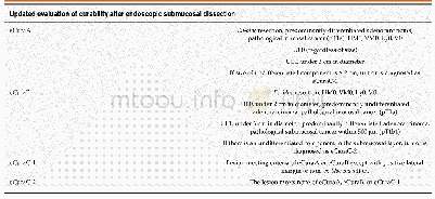
- Table 3 Updated evaluation of curability after endoscopic submucosal dissection in Japanese gastric cancer treatment gui


