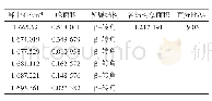
《Table 2.XRD data for the three most intense peaks of gibbsite》
 提示:宽带有限、当前游客访问压缩模式
本系列图表出处文件名:随高清版一同展现
《Mineral structure and crystal morphologies of high-iron hydrargillite》
The diffraction data of gibbsite and the corresponding standard card data are shown in Table 2.As shown in Table2,the lower relative intensities of the(110)and(200)peaks indicate that the sample is poorly crystalline.The interplanar spacings of the(002)and(110)planes are almost the same as those reported in the standard card.The(200)interplanar distance of the sample is smaller,which,in conjunction with the 2θshift,suggests that the crystal unit cell of the gibbsite in hydrargillite is smaller than that of standard gibbsite.
| 图表编号 | XD002866200 严禁用于非法目的 |
|---|---|
| 绘制时间 | 2018.05.01 |
| 作者 | Hui-bin Yang、Feng-qin Liu、Xiao-lin Pan |
| 绘制单位 | Hangzhou Yinsheng Enterprise Management Co.,Ltd.、School of Metallurgical and Ecological Engineering,University of Science and Technology Beijing、School of Metallurgy,Northeastern University |
| 更多格式 | 高清、无水印(增值服务) |


