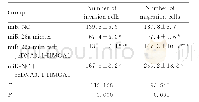
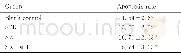
《表3 共转染后各组侵袭细胞数Tab.3 Number of invasion cells in various groups after co-transfection》
 提示:宽带有限、当前游客访问压缩模式
本系列图表出处文件名:随高清版一同展现
《miR-26a靶向HMGA1基因对结肠癌细胞生长、侵袭和迁移的影响》
*P<0.01 vs miR-NC group;△P<0.01 vs miR-26a mimics group;#P<0.01 vs miR-NC+pcDNA3.1-HMGA1group.
miR-NC组、miR-26a mimics组、miR-26a mimics+pcDNA3.1-HMGA1组和miR-NC+pcDNA3.1-HMGA1组细胞转染72h后,Transwell小室法检测细胞侵袭结果显示:miR-26amimics组和miR-26amimics+pcDNA3.1-HMGA1组侵袭细胞数均明显低于miR-NC组(t1=12.953,P1=0.000;t2=4.741,P2=0.002),miR-NC+pcDNA3.1-HMGA1组侵袭细胞数明显高于miR-NC组(t=4.956,P=0.001);miR-26a mimics+pcDNA3.1-HMGA1组侵袭细胞数明显高于miR-26amimics组(t=8.212,P=0.000),低于miR-NC+pcDNA3.1-HMGA1组(t=9.697,P=0.000)。见表3。
| 图表编号 | XD004407800 严禁用于非法目的 |
|---|---|
| 绘制时间 | 2018.11.28 |
| 作者 | 王廷刚、薛峰、李宇、牛兆健、王野 |
| 绘制单位 | 青岛海慈医疗集团普外科、青岛大学医学院附属医院普外科、青岛大学医学院附属医院普外科、青岛大学医学院附属医院普外科、青岛海慈医疗集团普外科 |
| 更多格式 | 高清、无水印(增值服务) |

