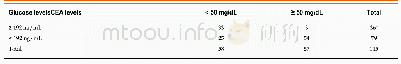
《Table 3 Correlation between human leukocyte antigen F-associated transcript 10 and p53 expression i
 提示:宽带有限、当前游客访问压缩模式
本系列图表出处文件名:随高清版一同展现
《Clinicopathological significance of human leukocyte antigen F-associated transcript 10 expression in colorectal cancer》
r=0.568,P=0.000.FAT10:Human leukocyte antigen F-associated transcript 10.
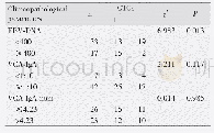
Immunohistochemical staining showed that positive signals,most of which were weak,were present only in four(6.56%)normal colorectal mucosal tissues and in 11(18.03%)tumor-adjacent tissues(Figure 1A).According to IS,only one(1.64%)normal colorectal mucosal tissue and six(9.84%)tumor-adjacent tissues were positive for FAT10.In contrast,46(75.41%)CRC tissues were positive for FAT10,of which 39showed moderately to strongly positive expression(Figure 1B).FAT10 expression was significantly higher in CRC than in normal colorectal mucosa and tumor-adjacent tissues(P<0.05),although there was no significant difference between normal colorectal mucosa and tumor-adjacent tissues(P>0.05;Table 1 and Figure 1C).
| 图表编号 | XD0043288300 严禁用于非法目的 |
|---|---|
| 绘制时间 | 2019.01.15 |
| 作者 | Chun-Yang Zhang、Jie Sun、Xing Wang、Cui-Fang Wang、Xian-Dong Zeng |
| 绘制单位 | Department of Emergency Medicine, Central Hospital Affiliated to Shenyang Medical College、Department of Pathology, Central Hospital Affiliated to Shenyang Medical College、Department of Pathology, Central Hospital Affiliated to Shenyang Medical College、Dep |
| 更多格式 | 高清、无水印(增值服务) |


