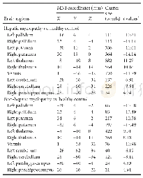
《Table 3 Z-statistical maps of gray matter volume differences among patients and normal controls》
 提示:宽带有限、当前游客访问压缩模式
本系列图表出处文件名:随高清版一同展现
《Microstructural damage pattern of vascular cognitive impairment: a comparison between moyamoya disease and cerebrovascular atherosclerotic disease》
x,y,z:Peak MNI coordinates;Z:statistical value for the voxel showing peak gray matter volume differences among groups(adjusted for age and sex,corrected P<0.05/3).MNI:Montreal Neurological Institute;NC:normal control;MMD:moyamoya disease;CAD:cerebrova
Similarly,we found that FA was significantly lower and MD was significantly higher in the central part of the white matter tracts in the cerebrovascular atherosclerotic disease group compared with controls,including a portion of the genu and body of corpus callosum,cingulum,posterior thalamic radiation,anterior corona radiata,and part of superior corona radiata.Higher MD was more extensive in these same white matter tracts.In addition,higher MD was also found in the bilateral internal and external capsule of the cerebrovascular atherosclerotic disease group.FA was not higher in the cerebrovascular atherosclerotic disease group than in controls,nor was MD lower(Tables 4 and 5).
| 图表编号 | XD0040614900 严禁用于非法目的 |
|---|---|
| 绘制时间 | 2019.05.01 |
| 作者 | Jia-Bin Su、Si-Da Xi、Shu-Yi Zhou、Xin Zhang、Shen-Hong Jiang、Bin Xu、Liang Chen、Yu Lei、Chao Gao、Yu-Xiang Gu |
| 绘制单位 | Department of Neurosurgery, Huashan Hospital, Fudan University、Shanghai Medical College, Fudan University、Department of Radiology, Huashan Hospital, Fudan University、Department of Neurosurgery, Huashan Hospital, Fudan University、Department of Neurosurgery |
| 更多格式 | 高清、无水印(增值服务) |


