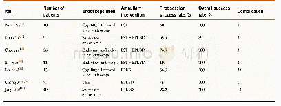
《Table 1 Transduodenal ampullectomy of ampullary lesions》
 提示:宽带有限、当前游客访问压缩模式
本系列图表出处文件名:随高清版一同展现
《Surgical method choice and coincidence rate of pathological diagnoses in transduodenal ampullectomy: A retrospective case series study and review of the literature》
1:Tubulovillous adenoma;2:Adenocarcinoma;3:Dysplasia;4:Adenomatous hyperplasia;5:Serrated adenoma;6:Intraepithelial neoplasia.M:Male;F:Female;y:Year;m:Month;pTis:Pathological carcinoma in situ;pT1:Pathological tumor limited to the ampulla of Vater or the
From December 2011 to October 2017,ten patients(four men and six women)with an average age of 66.2 years(range,51-77 years)underwent TDA for ampullary neoplasms at our hospital.Endoscopic biopsy results showed benign lesions(Table 1).Main clinical symptoms were jaundice and abdominal pain.All cases underwent a duodenal papilla biopsy(Figure 1A and B)and an intraoperative frozen-section pathological examination.All patients underwent EUS,computed tomography(CT),or magnetic resonance imaging(MRI)prior to surgery(Figure 2A-D)as well as preoperative examination,which indicated that there was no hepatic or pulmonary metastasis or abdominal lymph node enlargement.This study was approved by the ethics committee of our hospital,and all patients provided informed consent to participation in the study.This study was a single-center nonconsecutive retrospective case series study,and the work was reported in accordance with the PROCESS criteria[17].
| 图表编号 | XD0054204000 严禁用于非法目的 |
|---|---|
| 绘制时间 | 2019.03.26 |
| 作者 | Feng Liu、Jia-Lin Cheng、Jing Cui、Zong-Zhen Xu、Zhen Fu、Ju Liu、Hu Tian |
| 绘制单位 | Department of General Surgery,Shandong Provincial Qianfoshan Hospital,Shandong University、Department of General Surgery,Shandong Provincial Qianfoshan Hospital,Shandong University、Taishan Medical University、Department of Pathology,Shandong Provincial Qian |
| 更多格式 | 高清、无水印(增值服务) |
查看“Table 1 Transduodenal ampullectomy of ampullary lesions”的人还看了
-

- Table 7 Success rates of stone removal in Billroth II reconstruction in different ampullary interventions
-

- Table 8 Diagnostic accuracy parameters for significant colonic lesion detection based on quantitative faecal immunochemi


