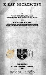
《X-RAY MICROSCOPY》
| 作者 | V.E. COSSLETT AND W.C. NIXON 编者 |
|---|---|
| 出版 | 未查询到或未知 |
| 参考页数 | |
| 出版时间 | 没有确切时间的资料 目录预览 |
| ISBN号 | 无 — 求助条款 |
| PDF编号 | 820349628(仅供预览,未存储实际文件) |
| 求助格式 | 扫描PDF(若分多册发行,每次仅能受理1册) |
CHAPTER 1Introduction1
1.1.The use of X-rays for microscopy1
1.2.Contact microradiography2
1.3.Reflexion X-ray microscopy4
1.4.Point projection with X-rays7
1.5.Scanning methods9
1.6.Comparison with optical and electron microscopy9
1.6.1.Penetration and absorption in matter10
1.6.2.X-ray emission analysis11
1.6.3.Microdiffraction12
1.6.4.Stereomicro-scopy12
1.6.5.Resolving-power13
1.7.Scope and applications of X-ray microscopy14
1.7.1.Materials opaque to light14
1.7.2.Structure of thick specimens14
1.7.3.Living organisms14
1.7.4.Quantitative microanalysis in biology15
1.7.5.Quantitative microanalysis of inorganic specimens15
1.8.Other X-ray methods16
1.8.1.'Darkfield'imaging by the Berg-Barrett method16
1.8.2.Optical reconstruction of lattice structure17
1.8.3.Diffraction microscopy17
CHAPTER 2Contact Microradiography19
2.1.Introduction19
2.2.Absorption of X-rays in matter21
2.2.1.Absorption coefficients22
2.2.2.Fluorescence radiation25
2.2.3.Absorption edges26
2.3.Optimum conditions for microradiography27
2.3.1.Wavelength and specimen thickness28
2.3.2.Choice of characteristic,continuous or fluorescent radiation33
2.4.Geometrical blurring of the micro-image34
2.5.Photographic and optical requirements37
2.5.1.High resolution emulsions37
2.5.2.Optical enlarging system40
2.6.Ultimate resolving-power of the contact method40
2.6.1.Unsharpness in image formation41
2.6.2.Image spread41
2.6.3.Size and aggregation of photographic grains42
2.6.4.Resolving-power of the enlarging system43
2.6.5.Contrast requirements43
2.6.6.Ultimate resolution43
2.7.Associated techniques of microradiography44
2.7.1.Stereo-microradiography44
2.7.2.Microfluoroscopy45
2.7.3.Electron microradiography45
CHAPTER 3Microscopy by Point Projection48
3.1.Introduction48
3.2.The geometry of projection50
3.3.Fresnel diffraction54
3.4.Experimental systems58
3.4.1.Camera obscura58
3.4.2.Point anode tube59
3.4.3.Window target tube60
3.5.Electron optical limitations62
3.5.1.Cathode emission62
3.5.2.Lens aberrations63
3.6.Target limitations66
3.6.1.Production and absorption of X-rays66
3.6.2.Thermal dissipation67
3.6.3.Electron scattering68
3.6.4.Choice of target material71
3.7.Photographing the image72
3.8.Comparison of the contact and projection methods74
3.8.1.Resolution74
3.8.3.Field of view74
3.8.3.Exposure-time77
3.8.4.Experimental convenience78
3.9.Intermediate magnifications80
3.9.1.Arbitrary magnification81
3.9.2.The times-two method84
CHAPTER 4Reflexion X-Ray Microscopy:Mirror Systems87
4.1.Total reflexion of X-rays87
4.2.Dioptric focusing91
4.3.Focusing by total reflexion94
4.4.Aberrations of a cylindrical mirror of circular section98
4.4.1.Spherical aberration100
4.4.2.Coma102
4.4.3.Obliquity of the field103
4.4.4.Curvature of the field104
4.5.Astigmatism and distortion105
4.6.Compound systems of circular mirrors109
4.7.Compound systems of figured mirrors111
4.8.Practical design of the reflexion X-ray microscope114
4.8.1.Experimental arrangements116
4.9.Practical limitations118
4.10.The ultimate limit set by diffraction120
CHAPTER 5Reflexion X-Ray Microscopy:Curved Crystals123
5.1.Efficiency of Bragg reflexion124
5.2.Focusing at glancing incidence126
5.2.1.Crystals curved at right angles to the plane of incidence127
5.3.Focusing at near normal incidence131
5.4.Focusing by ring mirrors136
CHAPTER 6X-Ray Absorption Microanalysis139
6.1.Microanalysis by differential absorption139
6.1.1.Measurements at an absorption edge142
6.1.2.Microweighing and microanalysis144
6.2.Dry weight determination145
6.2.1.Principles of the method145
6.2.2.Microphotometry147
6.2.3.Experimental errors149
6.2.4.Resolving-power and limit of detection150
6.2.5.Improvements in the contact method151
6.2.6.Water content152
6.3.Absorption micro-spectrometry by the contact method153
6.3.1.Procedure153
6.3.2.The spectrometer156
6.3.3.Experimental errors157
6.4.Photo-graphic considerations160
6.4.1.The characteristic curve160
6.4.2.Grain size and granularity163
6.4.3.Variation of sensitivity with wave-length165
6.4.4.Equivalent effective wavelength166
6.5.Absorption analysis by the projection method170
6.5.1.X-ray tube and spectrometer171
6.5.2.The recording system175
6.5.3.Errors in counter recording178
6.5.4.Comparison with the contact method181
CHAPTER 7X-Ray Emission Microanalysis183
7.1.Emission microspectrometry183
7.1.1.Experimental conditions for direct excitation183
7.1.2.Corrections;limits of accuracy188
7.1.3.Discrimination between elements193
7.1.4.Comparison with other methods196
7.2.The static spot microanalyser197
7.3.The scanning microanalyser or flying-spot X-ray microscope202
7.3.1.Apparatus202
7.3.2.Applications205
7.4.Fluorescent microspectrometry206
CHAPTER 8Production of X-Rays212
8.1.Electron emission and focusing212
8.1.1.General requirements212
8.1.2.Thermionic emission214
8.1.3.Field emission215
8.2.Heat dissipation in the target217
8.2.1.Thick target217
8.2.2.Thin target217
8.2.3.Needle target219
8.2.4.Heat balance in a thin target220
8.3.Electron range in the target222
8.4.Spectral distribution223
8.5.Efficiency of X-ray production226
8.5.1.The continuous spectrum226
8.5.2.The characteristic spectrum228
8.6.Angular distribution232
8.7.Filtration and other means of monochromatization236
8.7.1.Filters236
8.7.2.Self-filtration237
8.7.3.Total reflexion238
8.7.4.Crystal monochromators240
CHAPTER 9Specimen Preparation Techniques241
9.1.Biological and medical specimens241
9.1.1.Freeze-drying241
9.1.2.Embedding and sectioning242
9.1.3.Injection and staining244
9.1.4.Mounting the specimen for contact microradiography246
9.1.5.Mounting the specimen for projection or reflexion X-ray microscopy249
9.1.6.Living organisms249
9.2.Metallurgical and industrial specimens250
9.2.1.Sectioning by grinding251
9.2.2.Shadow-casting and replica methods252
CHAPTER 10Techniques of Contact Microradiography254
10.1.Apparatus for contact microradiography254
10.1.1.General considerations254
10.1.2.Apparatus for hard radiation(<2?)256
10.1.3.Apparatus for soft radiation(2-10?)258
10.1.4.Apparatus for ultra-soft radiation(>10?)260
10.2.Recording the microradiograph263
10.2.1.Photographic considerations263
10.2.2.Optical enlargement263
10.3.Stereoscopic microradiography264
10.4.Microfluoroscopy266
CHAPTER 11Techniques of Projection Microscopy270
11.1.The electron gun270
11.2.The focusing system272
11.2.1.Single-stage demagnification273
11.2.2.Two-stage demagnification273
11.2.3.Astigmatism and its correction276
11.2.4.Comparison of electro-static and magnetic lenses279
11.3.Constructional details281
11.3.1.Magnetic lens model281
11.3.2.Electrostatic lenses284
11.4.Mechanical and electrical stability285
11.5.Electrical supplies288
11.6.Vacuum requirements289
11.7.Microscope alinement291
11.8.Microscope focusing292
11.9.Photographic recording296
11.10.Stereographic projection296
11.11.Transmission targets300
11.12.Maintenance of projection X-ray microscopes303
11.13.Commercial models of the projection microscope304
CHAPTER 12Applications of X-Ray Microscopy in Biology and Medicine306
12.1.Living specimens306
12.1.1.Rabbit306
12.2.1.Insects308
12.1.3.Ciné X-ray microscopy308
12.1.4.Radiation dosage to the specimen309
12.2.Thick sections310
12.3.Thin sections314
12.4.Botanical specimens317
12.5.Quantitative applications319
CHAPTER 13Inorganic Applications of X-Ray Microscopy322
13.1.Metallurgy322
13.1.1.Iron and steel-segregations and inclusions322
13.1.2.Cast iron323
13.1.3.Iron ores and sinters324
13.1.4.Other ores and minerals325
13.1.5.Light metal alloys325
13.1.6.Diffusion328
13.1.7.Spot welding329
13.1.8.Castings329
13.1.9.Other metallurgical applications330
13.2.Industrial applications331
13.2.1.Paper331
13.2.2.Fibres and fabrics332
13.2.3.Geology333
13.2.4.Miscellaneous334
CHAPTER 14Microdiffraction336
14.1.X-ray tubes336
14.1.1.Micro-focus X-ray tubes337
14.1.2.Reflexion types340
14.2.Cameras and methods342
14.2.1.Transmission344
14.2.2.Back reflexion344
14.2.3.Forward reflexion345
14.2.4.Divergent beam347
14.2.5.Selected area349
14.2.6.Powder cameras349
14.2.7.Moving specimen and film types350
14.3.Conclusion350
CHAPTER 15Some New Experimental Methods351
15.1.The scanning system as an X-ray image intensifier351
15.2.Other forms of image intensifier354
15.3.Image conversion X-ray microscope356
15.4.Projection X-ray microscopy:focusing by back-scattered electrons359
15.5.Projection X-ray microscopy:focusing by forward-scattered electrons362
15.6.Contact microradiography with electron microscope enlargement365
COMMENTS ADDED IN PROOF367
APPENDIXAbsorption and Emission Data372
A.1.X-ray mass-absorption coefficients of certain elements and of dry air for the wavelength range 0.710-8.321 ?372
A.2.X-ray mass absorption coefficients of certain elements for the wavelength range 8.34-44 ?375
A.3.Principal emission lines of the K-series(wavelengths in ? units)375
A.4.Principal emission lines of the L-series(wavelengths in ? units)377
REFERENCES380
INDEX403
《X-RAY MICROSCOPY》由于是年代较久的资料都绝版了,几乎不可能购买到实物。如果大家为了学习确实需要,可向博主求助其电子版PDF文件。对合法合规的求助,我会当即受理并将下载地址发送给你。
高度相关资料
-

- X-RAY METHODS
- JOHN WILEY & SONS
-

- X-RAY METALLOGRAPHY
- JOHN WILEY AND SONS
-

- COSMIC X-RAY ASTRONOMY
- 1980 ADAM HILGER LTD
-
- X-RAY OPTICS
- 1949 METHUEN & CO LTD
-

- Better X-Ray Interpretation
- 1997 Springhouse Pub Co
-

- X-RAY CRYSTALLOGRAPHY
- 1970 CAMBRIDGE UNIVERSITY PRESS
-

- X-RAY DIFFRACTION
- 1969 ADDISON-WESLEY PUBLISHING COMPANY
-

- DISLOCATION STUDIES IN DIAMOND BY X-RAY DIFFRACTION MICROSCOPY
- 1963 DEPT. OF COMMERCE
-

- SUPERVOLTAGE X-RAY THERAPY
- 1944 H.K.LEWIS & CO.LTD
-
- SCANNING ELECTRON MICROSCOPY AND X-RAY MICROANALYSIS SECOND EDITION
- 1992 PLENUM PRESS . NEW YORK AND LONDON
提示:百度云已更名为百度网盘(百度盘),天翼云盘、微盘下载地址……暂未提供。➥ PDF文字可复制化或转WORD
