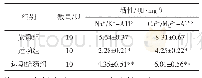
《表2 各组大鼠咬肌细胞外Na+浓度Tab.2 Extracellular Na+concentration in the masseter muscle of the rats at differe
 提示:宽带有限、当前游客访问压缩模式
本系列图表出处文件名:随高清版一同展现
《咬合干扰大鼠咬肌线粒体钙超载的离子变化特征及其钙调蛋白激酶Ⅱ的调节机制》
aP<0.05 vs control on the same side;bP<0.05 vs the left side in the same group.
根据分光光度计测得的A值带入公式计算,实验组咬合干扰侧咬肌细胞外Na+浓度在建模3 d组、7 d组、14 d组及21 d组呈持续升高趋势(P<0.05),去除咬合干扰后3 d组细胞外Na+浓度显著降低(P<0.05),实验组非干扰侧变化趋势同干扰侧但略低于干扰侧(P<0.05,表2)。
| 图表编号 | XD0016636800 严禁用于非法目的 |
|---|---|
| 绘制时间 | 2018.06.20 |
| 作者 | 曾林、刘静 |
| 绘制单位 | 暨南大学口腔医学院、华侨医院口腔科、暨南大学口腔医学院修复学教研室 |
| 更多格式 | 高清、无水印(增值服务) |


